

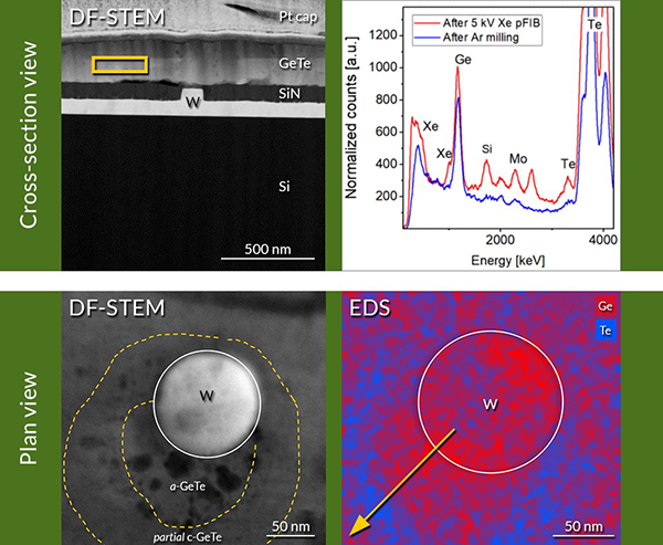
TEM specimens of GeTe-based, mushroom-type phase change memory (PCM) devices were prepared using a Xe plasma FIB tool. Xe plasma FIB (pFIB) preparation would be the preferred preparation technique over Ga FIB because the phase change layer, GeTe, is susceptible to ion beam damage. However, producing electron-transparent TEM specimens using a Xe pFIB is tedious and deviates from the standard Ga FIB method because of the relatively large Xe beam and its wide beam tails [1]. To obtain high-quality specimens free from Xe damage and suitable for high-resolution imaging and analysis, controlled specimen thinning of the cross-section and plan view specimens is achieved by post-pFIB polishing by concentrated Ar ion beam milling using Fischione Instruments’ Model 1080 PicoMill® TEM specimen preparation system.
Click here to view the webinar.
Click here to read the full document.
To speak with one of our Sales & Applications Engineers please call 01582 764334 or click here to email.
Lambda Photometrics is the leading UK Distributor of Characterisation, Measurement and Analysis solutions with particular expertise in Instrumentation, Laser & Light based products, Optics, Electro-optic Testing, Spectroscopy, Machine Vision, Optical Metrology, Fibre Optics, Microscopy and Anti-vibration tables & custom solutions
-

Fischione Model 1080 PicoMill® TEM Specimen Preparation System
- Achieve ultimate specimen quality – free from amorphous and implanted layers
- Complements FIB technology
- Milling without introduction of artifacts
- Advanced detector technology for imaging and precise endpoint detection
- In situ imaging with ions and electrons
- Microscope connectivity for risk-free specimen handling
- Adds capability and capacity
- Fast, reliable and easy to use